What is it about?
We removed 40% of the tip of mouse salivary glands to study how the glands respond to injury. While most of the salivary gland recovered from this injury with no apparent detrimental effects on the ability of cells to produce saliva, one region of the gland sustained a scar that contained reduced numbers of saliva-producing cells. We used RNA profiling to examine the injury response at 3 and 14 days after the injury. Three days after injury, most of the gland responded with activation of molecules known to be activated with wound repair. By 14 days after the injury, the injury response was almost complete as the gland became similar to non-injured glands. However, in the abnormal region with reduced saliva-producing cells, there was a persistent scar that included increased numbers of macrophages, which are immune cells that are known to stimulate scarring and participate in the control of inflammation. The scarred region included part, but not all, of the cut edge and persisted in the glands for up to 56 days, suggesting that the scarred region is a persistent fibrotic region.
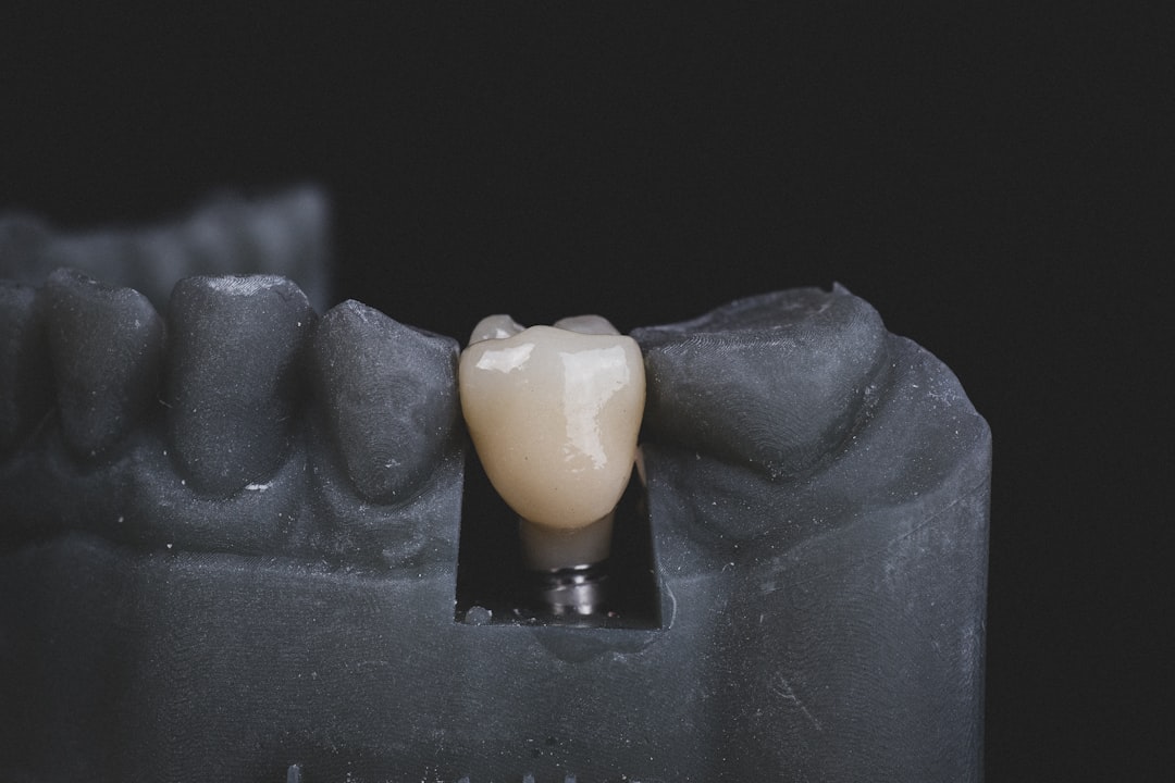
Featured Image

Photo by Akshay Nanavati on Unsplash
Why is it important?
Scarring is also known as fibrosis, which is a causative factor in loss of organ function in many organs, and there are few effective therapeutics available to reduce fibrosis. The partially resected salivary gland model is useful model to study why some parts of the gland recover from injury with an apparent maintenance of function and why other parts become fibrotic. There is a lot of regional heterogeneity in the salivary glands of patients that have the autoimmune disease, Sjogren's Syndrome, and produce very low levels of saliva. This model may also eventually help us to understand heterogeneity in patients.
Perspectives
This is the first publication using an in vivo manipulation by the Larsen lab. This project was a huge effort that involved many people, primarily the collaborative efforts of three graduate students. We hope that in future work that we can figure out why the different regions of the gland respond differently and that this will provide some insight into development of new therapeutics for human diseases.
Dr. Melinda Larsen
University at Albany, State University of New York
Read the Original
This page is a summary of: Regional Differences following Partial Salivary Gland Resection, Journal of Dental Research, November 2019, SAGE Publications,
DOI: 10.1177/0022034519889026.
You can read the full text:
Resources
Contributors
The following have contributed to this page


