What is it about?
By measuring spontaneous neural activity from neurons in higher order visual areas and mapping their whole brain relationship using fMRI, we have shown that a single neuron has restricted and circumscribed functional connections with different brain regions. Interestingly part of these associations was not only restricted to visual areas but to neuromodulatory centers as well.
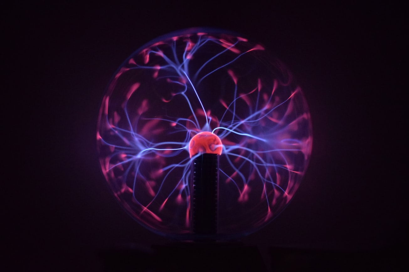
Featured Image

Photo by Stefano Bucciarelli on Unsplash
Why is it important?
These findings demonstrate that the activity of neurons is selectively coupled to discrete brain regions, with the coupling governed in part by anatomical network connections and also by indirect neuromodulatory ascending pathways.
Perspectives
Writing and working in this manuscript was a great pleasure as it taught me so much about the brain's self dynamics and association with distinct brain regions. It is also a continuation on my research interest and future goals in understanding how the influx of information (e.g. sensory information) may change depending on brain state and cognitive influence. I hope you find this article interesting
Daniel Zaldivar
National Institute of Mental Health
Read the Original
This page is a summary of: Brain-wide functional connectivity of face patch neurons during rest, Proceedings of the National Academy of Sciences, August 2022, Proceedings of the National Academy of Sciences,
DOI: 10.1073/pnas.2206559119.
You can read the full text:
Contributors
The following have contributed to this page


