What is it about?
The research presented in this paper demonstrates a new methodology that makes possible imaging biological material (proteins in the example shown), while simultaneously providing the spectroscopic chemical characterization via Infrared spectra of biomaterials in vitro, i.e., in their natural biological medium, and follow their chemical and physical evolution. The method described combines atomic force microscopy with infrared illumination of the sharp tip of the microscopy, together with graphene capped cells, achieving a high resolution of 10-25 nm in x,y, and z. This provides biologist and material scientists an excellent tool to unravel structure and function of soft material like polymers and biomaterials at the most fundamental level.
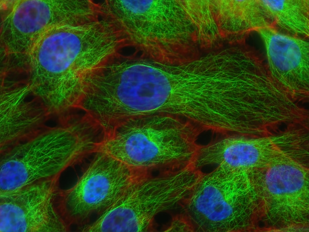
Featured Image
Photo by National Cancer Institute on Unsplash
Why is it important?
The growing research in bioengineering calls for in vitro, non-invasive nanoscale characterization of biological macromolecules. However, current imaging tools often use ionizing radiation under high vacuum and/or cold temperature, conditions that are far from the native biological environment. Our graphene-based platform combined with AFM and nano-FTIR makes opens the way for in vitro studies of the structural and chemical evolution of biological material, exemplified here by the study of the self-assembly of SbpA proteins on graphene. We have shown that the dynamical evolution of the protein substructure depends on their environment, the presence of ions such as Ca2+, protein-substrate and protein-water interactions.
Perspectives
The new methodology described in this article offers new solutions to operando characterization of proteins. Our new approach is less invasive, and avoids damage caused by radiation. The nanoscale structure information will provide new fundamental knowledge of protein assembly. We also believe that our platform opens opportunities for functional studies of other soft materials, ranging from biomaterial structures (ribosome, virus) to plastic polymer material, under in vitro conditions and external stimuli. We can foresee improvements in the technique to increase the resolution to below 10 nm, as this depends on the radius of the Atomic Force Microscopy.
Xiao Zhao
Read the Original
This page is a summary of: In vitro investigation of protein assembly by combined microscopy and infrared spectroscopy at the nanometer scale, Proceedings of the National Academy of Sciences, August 2022, Proceedings of the National Academy of Sciences,
DOI: 10.1073/pnas.2200019119.
You can read the full text:
Contributors
The following have contributed to this page