What is it about?
We mapped changes in the distribution of synapses in the larval zebrafish brain that occur with associative memory formation. We found that memory formation is associated with intense synapse proliferation in one area of the dorsal pallium (homolog of the amygdala and hippocampus) and synapse loss in another region. Neurons in the area of synapse proliferation became active upon memory recall. We observed only small, random changes in synapse strength that were not distinguishable from the background.
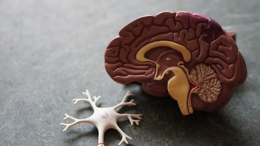
Featured Image

Photo by Hal Gatewood on Unsplash
Why is it important?
This is the first time that synapse change has been mapped during associative memory formation by longitudinal imaging. It establishes the anterolateral dorsal pallium as a likely locus of associative memory storage in the zebrafish. Memory formation appears to be associated with region-specific synapse loss and gain but changes in synapse strength were not observed. This may help to explain why associative memories are so robust.
Perspectives
This study illustrates the power of combining diverse technologies - recombinant FingR probes for labeling synapses, a custom-built light sheet microscope optimized for imaging small, faint signals (Scott Fraser and Thai Truong), and a system for recording, analyzing, and storing data that maximizes reproducibility and transparency (Carl Kesselman and Karl Czajkowski).
Don Arnold
University of Southern California
Read the Original
This page is a summary of: Regional synapse gain and loss accompany memory formation in larval zebrafish, Proceedings of the National Academy of Sciences, January 2022, Proceedings of the National Academy of Sciences,
DOI: 10.1073/pnas.2107661119.
You can read the full text:
Contributors
The following have contributed to this page