What is it about?
Most joint imaging technologies identify structural changes in the bone (x-rays) or soft tissues (MRI), which show how the sum of damage over time affected the tissue structure, but they do not show what is going on in the present. Here, we image the activity of enzymes in a living mouse knees, which provides real-time insight into the processes that will later result in structural changes. We also characterize the imaging reagents for quantifying changes in explant cartilage injury models.
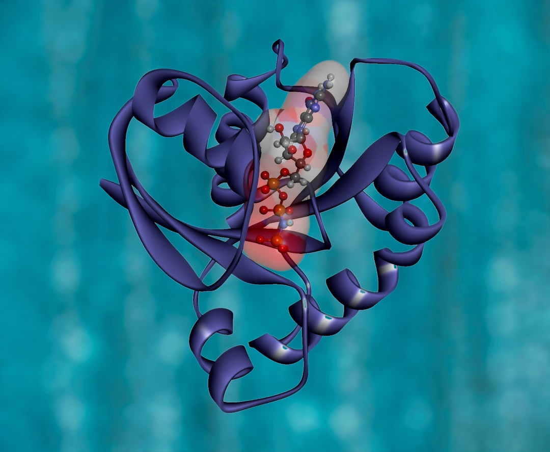
Featured Image
Why is it important?
Real time imaging of degradative processes allows for more rapid evaluation of therapeutic intervention strategies.
Read the Original
This page is a summary of: In-vitro and in-vivo imaging of MMP activity in cartilage and joint injury, Biochemical and Biophysical Research Communications, May 2015, Elsevier,
DOI: 10.1016/j.bbrc.2015.03.100.
You can read the full text:
Contributors
The following have contributed to this page