What is it about?
In the present study, we utilized autometallography (AMG) to localize the Ag in the liver and kidney tissues of cetaceans and developed a model called the cetacean histological Ag assay (CHAA) to estimate the Ag concentrations in the liver and kidney tissues of cetaceans.
Featured Image

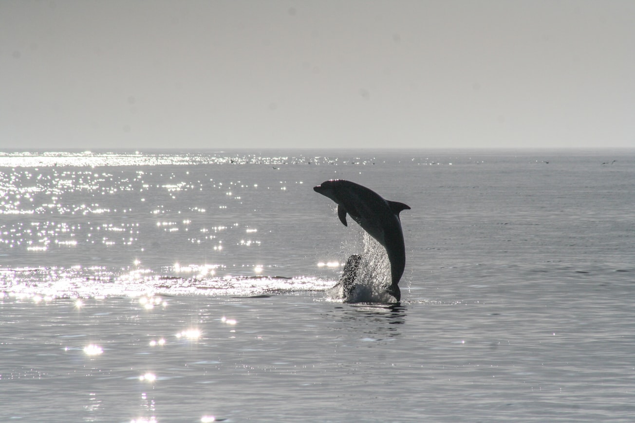
Photo by Noah Boyer on Unsplash
Why is it important?
Our results revealed that Ag was mainly located in hepatocytes, Kupffer cells and the epithelial cells of some proximal renal tubules. The tissue pattern of Ag/Ag compounds deposition in cetaceans was different from those in previous studies conducted on laboratory rats. This difference may suggest that cetaceans have a different metabolic profile of Ag, so a presumptive metabolic pathway of Ag in cetaceans is advanced. Furthermore, our results suggest that the Ag contamination in cetaceans living in the North-western Pacific Ocean is more severe than that in cetaceans living in other marine regions of the world. The level of Ag deposition in cetaceans living in the former area may have caused negative impacts on their health condition.
Perspectives
Further investigations are warranted to study the systemic Ag distribution, the cause of death/stranding, and the infectious diseases in stranded cetaceans with different Ag concentrations for comprehensively evaluating the negative health effects caused by Ag in cetaceans.
Dr. Wen-Ta Li
Fishhead Labs LLC
Read the Original
This page is a summary of: Investigation of silver (Ag) deposition in tissues from stranded cetaceans by autometallography (AMG), Environmental Pollution, April 2018, Elsevier,
DOI: 10.1016/j.envpol.2018.01.010.
You can read the full text:
Resources
Contributors
The following have contributed to this page


