What is it about?
A protocol is presented to localize Ag in cetacean liver and kidney tissues by autometallography. Furthermore, a new assay, named the cetacean histological Ag assay (CHAA) is developed to estimate the Ag concentrations in those tissues.
Featured Image

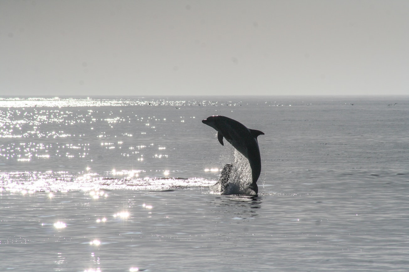
Photo by Noah Boyer on Unsplash
Why is it important?
The assay for estimating the Ag concentration is named the Cetacean Histological Ag Assay (CHAA), which is a regression model established by the data from image quantitative analysis of the AMG method and ICP-MS. The use of AMG with CHAA to localize and semi-quantify heavy metals provides a convenient methodology for spatio-temporal and cross-species studies.
Perspectives
FFPE samples are relatively easy to collect and store, and our previous study has demonstrated that the current AMG method can successfully amplify FFPE samples stored for over 15 years12. The mechanism of AMG is not affected by different animal species, for it has been wildly used in various animal species. Although the current article is focused on the cetaceans, the protocols described here may also be used in different animal species. In addition, the cost of the AMG method with ICP-MS is relatively low (as compared to laser ablation-ICP-MS), and thus the current protocols are valuable for researchers or countries lacking sufficient research funding to investigate the distribution and concentration of heavy metals in animal tissues. In conclusion, the use of AMG with quantitative analysis to localize and semi-quantify heavy metals provides a convenient methodology for spatio-temporal and cross-species studies.
Dr. Wen-Ta Li
Fishhead Labs LLC
Read the Original
This page is a summary of: Use of Autometallography to Localize and Semi-Quantify Silver in Cetacean Tissues, Journal of Visualized Experiments, October 2018, MyJove Corporation,
DOI: 10.3791/58232.
You can read the full text:
Resources
Contributors
The following have contributed to this page


