What is it about?
This study aimed to assess the clinical efficiency of combined awake craniotomy with 3-T intraoperative MRI (iMRI)–guided resection of gliomas adjacent to eloquent cortex. It also sought to explore the contribution of iMRI to surgeons’ learning process of maximal safe resection of gliomas.
Featured Image

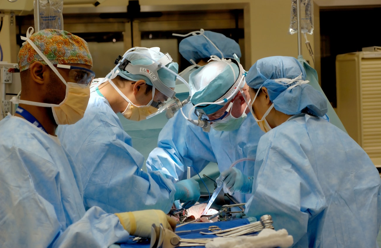
Photo by National Cancer Institute on Unsplash
Why is it important?
The analysis of one hundred six consecutive cases revealed that combined awake craniotomy and iMRI is a safe and efficient technique allowing maximal safe resection of eloquent area gliomas with possible subsequent benefit in terms of the overall and progression-free survival. Although there is a learning curve for applying this technique, it can also improve the surgeon’s ability in eloquent glioma surgery.
Perspectives
The synergistic use of combined intraoperative imaging and neurophysiological monitoring for gliomas located in proximity to eloquent areas are necessary when dealing with these challenging tumors. The neurophysiological testing and functional mapping used to delineate the cortico-subcortical functional boundaries need to be individualized and require extensive knowledge of the anatomical peculiarities of the tumor. This approach can allow an increase in the overall survival and quality of life for patients affected by these devastating tumors.
Dr. Cristina Diana Ghinda
Johns Hopkins University
Read the Original
This page is a summary of: Contribution of combined intraoperative electrophysiological investigation with 3-T intraoperative MRI for awake cerebral glioma surgery: comprehensive review of the clinical implications and radiological outcomes, Neurosurgical FOCUS, March 2016, Journal of Neurosurgery Publishing Group (JNSPG),
DOI: 10.3171/2015.12.focus15572.
You can read the full text:
Contributors
The following have contributed to this page


