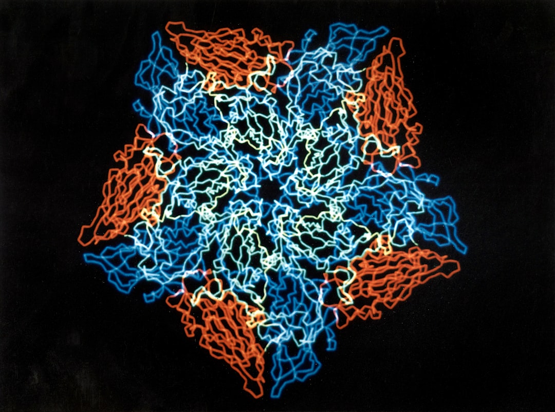
What is it about?
In Alzheimer’s disease patients, the protein amyloid-beta (Aβ) clumps up in the brain to form so-called fibrils. This has a toxic effect on the surrounding nerve cells. It is believed that immune cells compact the Aβ fibrils into what are known as plaques. It is now possible to track the development of these microscopically small structures in human using infrared microscopy.
Featured Image

Photo by Robina Weermeijer on Unsplash
Why is it important?
Aβ is the focus in the fight against Alzheimer’s disease. A central approach in the search for a cure is dissolving the plaques in the patients’ brains. The new findings indicate that the development of plaques could be stopped at an early stage by preventing the formation of oligomers. These are considered to be particularly harmful to the brain. The toxic effect of Aβ could thus be minimised with suitable drugs.
Perspectives
It has previously not been possible to directly observe the development of plaques. By combining methods from medicine and physics, new possibilities are now opening up. We are delighted to see that interdisciplinary approaches promote science as a whole!
Dominik Röhr
Ruhr-Universitat Bochum
Read the Original
This page is a summary of: Label-free vibrational imaging of different Aβ plaque types in Alzheimer’s disease reveals sequential events in plaque development, Acta Neuropathologica Communications, December 2020, Springer Science + Business Media,
DOI: 10.1186/s40478-020-01091-5.
You can read the full text:
Resources
Development of plaques in Alzheimer’s disease resolved
One cannot simply look inside someone else’s head. This makes it difficult to understand what is happening in the brains of people with dementia. A new method offers insights.
Drie manieren om naar alzheimer te kijken
Door drie imagingtechnieken te combineren, krijg je de groei van amyloïdeplaques ongekend helder in beeld. Het wijst misschien de weg naar moleculen die de progressie van de ziekte van Alzheimer kunnen afremmen.
Infrared imaing reveals the molecular composition of plaque.
On the left is a fully developed plaque of brown-stained Aβ. On the right, the structure of Aβ determined by infrared microscopy is shown. In core are fibrils (red), around are mainly oligomers (blue).
Contributors
The following have contributed to this page


