What is it about?
We present a case of a persistent left-sided inferior vena cava (IVC) affecting the side of approach in a patient undergoing lumbar interbody fusion through an oblique prepsoas retroperitoneal approach. Preoperative imaging of our patient revealed a persistent left-sided inferior vena cava with a wide interval between the aorta and the right-sided psoas, allowing us a right-sided oblique approach.
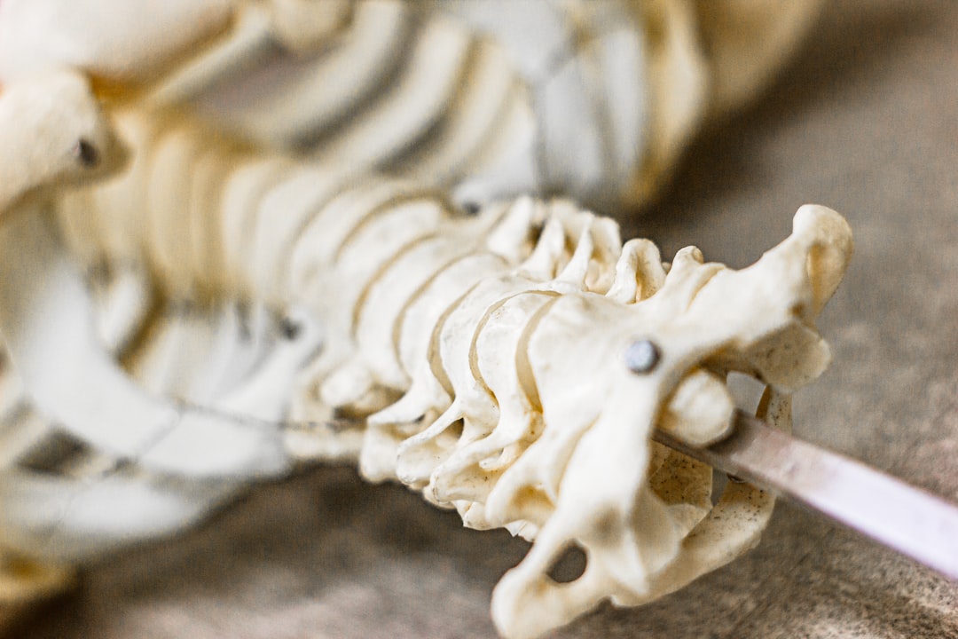
Featured Image

Photo by Olga Kononenko on Unsplash
Why is it important?
Thorough preoperative imaging evaluation is essential to identify vascular anomalies that may hinder oblique pre-psoas retroperitoneal approach to the lumbar spine. Although rare, double IVC or isolated Left IVC may complicate the oblique approach.
Perspectives
Based on our experience with this case and our review of the literature, we believe that such anomalies of the infra-renal IVC might best be visualized on coronal images. It is important to note that most MRI scans of the lumbar spine may not provide coronal sections anterior enough to assess the vascular anatomy. Evaluating all available axial MRI images going up to the thoracolumbar junction superiorly is important in such instances. Alternatively, coronal and axial images from abdominal CT scans may provide this information. Obtaining CT scans routinely for lumbar surgery carries the risk of unwarranted radiation exposure and is not something that we recommend. Evaluating prior abdominal CT scans of the patient, if available, can be a useful approach in such cases.
Dr Chirag A Berry
University of Cincinnati
Read the Original
This page is a summary of: Oblique Lumbar Interbody Fusion in Patient with Persistent Left-Sided Inferior Vena Cava: Case Report and Review of Literature, World Neurosurgery, December 2019, Elsevier,
DOI: 10.1016/j.wneu.2019.08.176.
You can read the full text:
Resources
Contributors
The following have contributed to this page


